CBCT (Cone Beam Computed Tomography)
is a widely used imaging tool in dentistry that allows us to create a more accurate three-dimensional image of the condition of the teeth compared to conventional X-ray images (panoramic, periapical). While conventional X-rays provide a two-dimensional projection of a three-dimensional image, with many layers overlapping, making it difficult to determine the relationship between anatomical structures. Panoramic X-rays also inevitably involve certain distortions. To avoid these uncertainties and potential inaccuracies, CBCT is increasingly being used.
How does CT work?
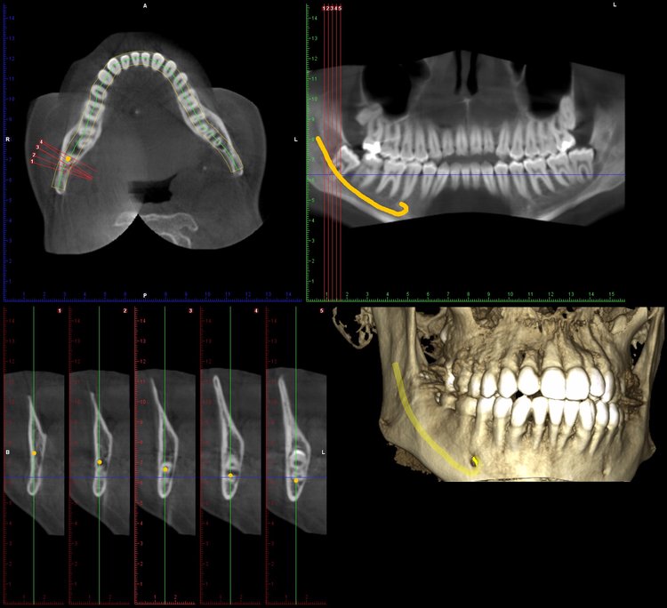
CBCT rotates around the patient’s head to capture images. The sensor registers these images, and then a program creates a series of axial slices from these images (primary reconstruction), which essentially contains the complete spatial mapping of the patient. Using this dataset, the software allows us to generate the images we use for diagnosis.
The image below shows the display of a CBCT scan of a dental arch:
When is CBCT advantageous?
In Oral Surgery:
Before the removal of wisdom teeth, some imaging procedure is necessary to determine the position of the tooth roots relative to major nerves within the jaw. While panoramic X-rays may not always precisely determine the root-nerve position, CBCT can provide an accurate view, assisting the oral surgeon in avoiding potential injuries.
In Endodontics:
Teeth roots have certain anatomical variations that may not always be visible on conventional periapical X-rays. These variations could include lateral or accessory canals or fractures. In certain cases, especially when inflammation affects the area around the tooth roots, traditional two-dimensional images may obscure the pathology. In the case of already root-filled teeth that remain symptomatic, a CBCT scan can be very useful. It allows for a sectional view of the filling, revealing any defects or untreated canals, enabling corrective measures.
In Implantology :
CBCT provides an accurate picture of the bone and soft tissues, making it widely used in implantology. It enables precise determination of bone availability with almost millimeter accuracy. This greatly facilitates the planning process for the dentist, as they can plan with precise certainty and see where bone augmentation may be necessary. Especially for surgeries involving the maxillary sinus or mandibular nerve, it provides excellent information about their precise morphology and any potential inflammation, aiding in avoiding injuries to important nerve structures.
Examples :
The example below illustrates that while the simple X-ray did not detect inflammation around the tooth root, it is clearly visible on the CT scan:
Panoramic X-ray:
CT scan:


